Showing posts with label microscopy. Show all posts

Monday, August 10, 2009

Mouse Perfusion and Dissection

I performed my first mouse perfusion today on a total of eight mice.
Perfusion is a special way to fix (preserve) an animal so that it can be later processed for electron microscopy. First, the mouse is anesthetized. Then, the chest cavity is opened up and a small incision is cut in the upper left side of the heart. A special needle is used to inject a saline solution into the lower right side of the heart. The saline flows through the mouse's entire circulatory system until it flows out of the opening made in the upper left side of the heart, completely replacing the blood. Finally, a syringe containing fixative replaces the one containing saline and the fixative is pumped into the mouse. When the perfusion is done, the mouse is stiff as a board. You can pick up dead mouse by it's completely straight tail resulting in what I call a, "mouse-sicle", or, "mouse-kabob."
After the mouse has been perfused properly, the necessary parts can be dissected. In this case, I dissected the intercostals (back rib muscles) and the leg muscles were dissected by another lab member.
A special thanks goes out the Hasna Baloui of the Salzer lab for teaching me the technique and letting me use her tools. Also, thank you Caterina Berti of the Burden lab for assisting me and dissecting the leg muscles.

Sunday, July 12, 2009

Tomography

About a month ago I posted an entry about the microscopes in my lab. In this previous post, I briefly described a technique called Electron Tomography. Here is great video that describes in detail the process of making a tomogram. The section used for this was 500 nanometers thick. This is extremely thick for an electron microscope. The data taken for this tomogram was gathered on one of the world's three 1 Million kV transmission electron microscopes! Most microscopes only have the capability to gather images on sections no thicker than 200 nanometers.

About a year ago I was in a class at the New York Structural Biology Center about electron microscopy techniques. The lesson about electron tomography was taught by the person who made this particular tomogram.

Tuesday, July 7, 2009

1950 RCA Tabletop Electron Microscope

Check out this beautiful example of Atomic Age design from RCA. This microscope was marketed as an affordable alternative to a typical Scanning Electron Microscope. "So simplified is the new instrument that a high school student or unskilled laboratory technician can quickly learn to use it!"
This is the cover from a twelve page brochure detailing the features of the RCA EMT Tabletop. The advertisement boasts a maximum magnification of 6,000x. Today's SEMs are capable of magnifications beyond 100,000x.

Monday, July 6, 2009

Carbon Atoms in Motion!

Here's some amazing footage of Carbon atoms moving around. The movie was captured with a Transmission Electron Microscope called TEAM 0.5. This microscope (cross-sectioned on the left) uses special lenses to correct for chromatic and spherical aberration, one of the limitations of a typical TEM. This is one of the key features that allows researchers to clearly view atoms and atomic lattices.
From the Article:
"Researchers with the U.S. Department of Energy's Lawrence Berkeley National Laboratory (Berkeley Lab), working with TEAM 0.5, the world's most powerful transmission electron microscope, have made a movie that shows in real-time carbon atoms repositioning themselves around the edge of a hole that was punched into a graphene sheet. Viewers can observe how chemical bonds break and form as the suddenly volatile atoms are driven to find a stable configuration. This is the first ever live recording of the dynamics of carbon atoms in graphene."

Sunday, June 28, 2009

Swine Flu Microscopy

Though I may be a little late blogging about this, H1N1 (swine flu) has some potential to stage a major comeback during the upcoming flu season. So, perhaps I'm early.
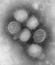
The image to the left of the H1N1 virus that was shown all over the news was taken on a Transmission Electron Microscope at the CDC Influenza Laboratory. The virus is about 80-120 nanometers in diameter. H1N1 was able to transmit from pigs to humans though what is called, "antigenic shifts." In fact, the H1H1 strain is a combination of bird influenza, swine influenza, and human influenza.
See this image and more like it in much higher resolution on the Centers for Disease Control and Prevention website.

Friday, June 26, 2009

The Nano Song

Here is the winner of the ACS-Nanonation's nano-video contest. If Jim Henson was around today, I think he'd approve.

...Nano!!!

Wednesday, June 24, 2009

Muscle fibers

dailymotion.com has some amazing videos made by a group called Weird_Weird_Science about zooming into objects. They zoom into hair, Aluminum, a tooth, concrete, and many others including this video of lice. In this video, the highlighted feature is muscle tissue.

Here (right)is image that I took on one of our microscopes (the Philips CM-12 TEM) of some muscle tissue from a mouse. This tissue looks better preserved than the lice muscle and may give you a better idea of how the muscle bundles are organized. Click to enlarge.

Thursday, June 18, 2009

More Amazing Antique Microscopes

Here is another site with some amazing images of antique microscopes. Allan Wissner of antique-microscopes.com has a massive collection of beautiful and impressive microscopes ranging from a 1770 Solar Microscope to a 1930 Carl Zeiss Spectroscope. If you're trying to find something specific, the site index has the collection organized by country of origin and maker. I could spend hours looking at these images. Speaking of which, a page of this site is dedicated to links of other microscope collections. Thank you Allan for your wonderful contribution to the microscopy world!

Tuesday, June 16, 2009

The Man in the White Suit

Here's a still from a neat 1951 film called, "The Man in the White Suit." Check out the clip. "..it's an electo..electron er..electron microscope."In the business, we call those EM-TJ's (Electron Microscopy Technician Jumpsuits). I wear mine everyday!

Molecular-Gear

Along the same lines as the molecular-car (something I would have blogged about had I started this four years ago) is the molecular-gear. This gear has a diameter of just 1.2 nanometers! The major breakthrough here is that this gear can be deliberately rotated. Previous developments in molecular gears resulted in random, uncontrolled rotation and displacement. Possible uses of this gear could include the building of molecular scale machinery for use in biological or material advances.
This was developed by a group (lead by Christian Joachim) from A*STAR’s Institute of Materials Research and Engineering (IMRE) in Singapore. You can download the full .pdf abstract here.

Sunday, June 14, 2009

Condenser Lens

Here is an interesting piece. This is a condenser lens. It is one of the most important parts of an electron microscope because controls the intensity of the electron beam.My boss gave this to me on one of my first days in the lab and it's one heck of a paperweight. The thing weighs a ton because it is essentially a giant coil of wire. The coil is a big controllable electromagnet and "condenses" the size of the electron beam as it passes through.

Thursday, June 11, 2009

Microscope Collection

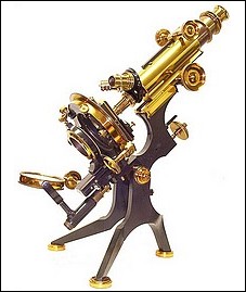
So, last week I talked about an amazing collection of microscopes at the University of Arizona College of Optical Sciences. This week, I decided to share my collection of microscopes with you. Again, they are not so much antique as they are vintage. The smaller one (right)was likely a model for school children in the 1940's or 50's. It came in a neat wooden box containing a few tools, a slide, and an old catalog from the company. Here (left) is an image I took with it of a piece of mica that i found in some potting soil.

The larger one (bottom right) with the odd looking bar is an old Carl Zeiss model (#316645 if you're interested). I consulted with a director of the Zeiss historical society about what it is and what it's function was and he thinks that it was measuring tool designed by Zeiss for use in the Zeiss factory for a quality.

Both of these microscopes were rather affordable. Probably because they were both extremely dirty when I bought them. They were obviously well used, especially the Zeiss. I spent several pleasant evenings in harsh light with a long drawn out whiskey, carefully removing every screw, opening every objective and condenser, polishing every lens, and loving every minute of it before putting it all back together. By the end, they look more or less brand new. In the Zeiss, I found a protractor in the eye-piece that I carefully aligned into the CORRECT position (the factory apparently neglected to do so in manufacturing). I also found a small prism in there.
Click the images to enlarge them.

Tuesday, June 9, 2009

Microscopes in the lab

Here are a couple of pictures I took of the two Transmission Electron Microscopes (click to enlarge) that are currently in our lab. I Photoshoped them a little to cut out the background. On the left is the Philips CM-12. This is our work-horse and the instrument that I use the most. It's source is a Tungsten Filament that can run at 120kV. Most of the TEM images that you will see on this blog were taken using this microscope.
On the right is the Philips CM-200. This microscope is mostly used by the structural biologists in the institute for studying protein crystals and single particles. It's source is a Field Emission Gun that can run at 200kV. The CM-200 has a special cryo-stage for doing cryo-electron microscopy can also be controlled by a computer. This is handy for doing Electron Tomography, a special type of imaging that can make 3D reconstructions. See movie below.

By the way, I can't take credit for this video. Whoever created it likely spent years developing getting the right conditions for the sample and many many hours on image acquisition and processing.

Sunday, June 7, 2009

Cancer Cells

Here's a neat video of some cancer cells imaged by the Cancer Research UK Electron Microscopy Unit, based at the London Research Institute.

Cancer cells are interesting. They don't communicate with other cells, don't seem to die off like normal cells (apoptosis), and they divide rapidly and invade the body. It is like the equivalent up having thousands of tiny foreign objects into your body.

Friday, June 5, 2009

Helium Ion Microscope

This week, we hosted a visit from Zeiss. They gave a presentation on a new technology that they just acquired called a Helium Ion Microscope. I remember seeing this demonstrated two years ago at the Microscopy & Microanalysis Convention in Chicago. Allis, which was purchased by Zeiss two years ago was the company that successfully figured out how to make it work.

This is a devise similar to a Scanning Electron Microscope, but uses Helium ions to create secondary electrons off the sample instead of an electron beam. This has advantages of getting much better resolution (due to it's incredibly small virtual source of three atoms), and an amazing depth of field compared to standard SEM. I think that this tool has the potential to replace the Scanning Electron Microscope, but they're about twice as expensive as a high-end SEM right now. There are only 7 being used in the world right now (soon to be 8). One is at Harvard and there is another in Singapore (where the speaker is teaching right now).
In all, the event was a success. Thirty plus people showed up from all over the New York area including people from Rockefeller University, the New York Structural Biology Center, and the New York University School of Medicine.

Thursday, June 4, 2009

Antique microscopes


I've recently begun to collect antique microscopes, but the two I own now would be considered more vintage than antique. I wish to one day own some like this from the University of Arizona College of Optical Sciences collection of antique microscopes.
There are some of the highest quality images of the most beautiful microscopes. Definitely worth a look if you are interested in antique microscope. They even have some made of wood from the 1700s.